


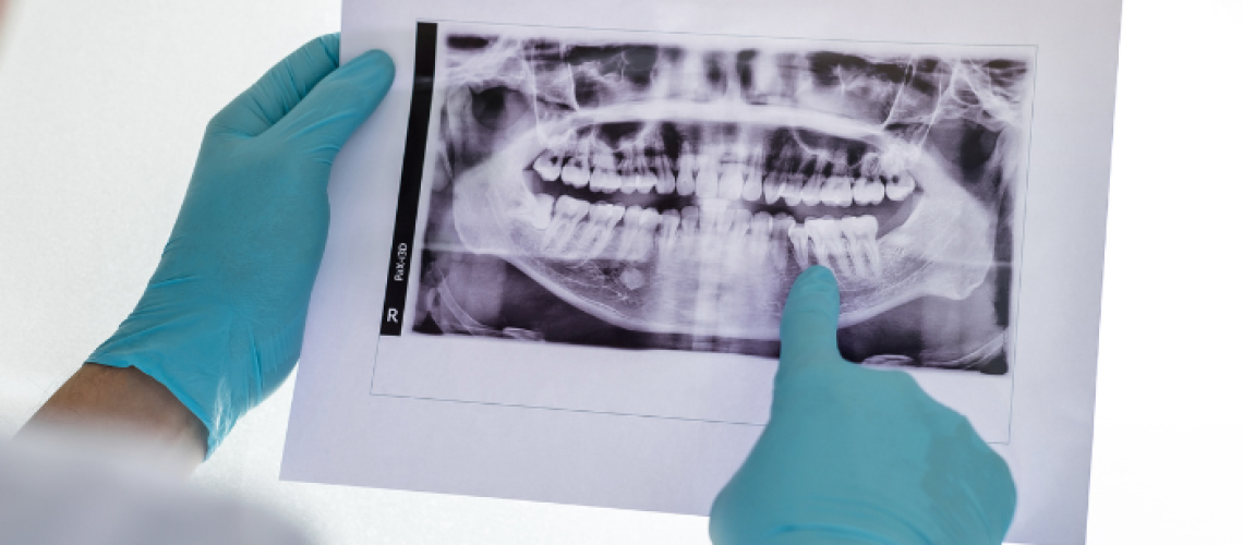

Many patients are surprised to learn that placing a dental implant without proper imaging can compromise both the safety and predictability of the implant procedure.
Before implant placement, detailed scans are essential to provide information about nerve locations, bone thickness, sinus positioning, and the overall structure of the jaw. These scans act as a precise roadmap, guiding the dentist on where an implant can be placed safely, how to avoid complications, and whether the existing bone can support the implant without additional preparation.
Even if replacing a missing tooth feels urgent, this diagnostic phase plays a critical role in ensuring safe and successful results.
In this article, we will explain the scans required during implant planning so you can move forward with confidence.
Why Traditional X-Rays Aren’t Enough
Traditional dental X-rays are useful for detecting cavities and evaluating general oral health, but they have clear limitations when it comes to implant planning.
Standard X-rays produce a flat, two-dimensional image. While they can confirm that bone is present, they do not provide enough information about its thickness, density, or relationship to nearby anatomical structures.
Planning an implant using only this type of image is similar to working without depth perception. At Kootenai Dental Group in Coeur d’Alene, ID, Dr. Delwyn Dick, DDS, carefully evaluates:
- Bone density, to determine whether the bone is strong enough to support an implant or if it requires reinforcement.
- Bone volume, to see the true width and depth of the available bone and ensure there is enough space for placement.
- Nerve locations, including the inferior alveolar nerve, to prevent permanent damage.
- Sinus proximity, to prevent sinus perforation. This is especially critical for implants in the upper jaw.
- Adjacent tooth roots, to prevent damage to neighboring healthy teeth.
Without 3D dental imaging, these details are less precise and more difficult to assess. Relying only on traditional X-rays may introduce additional risk and can lead to complications that are entirely preventable with modern technology.
What is a CBCT Scan?
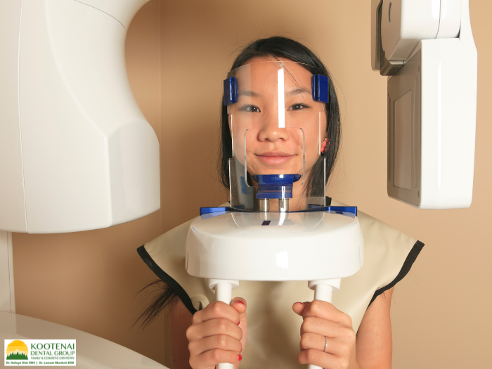
The current standard for dental implant planning is the Cone Beam Computed Tomography (CBCT) scan. This technology provides a comprehensive 3D view of the jaw and surrounding structures.
The scan usually takes only 10 to 20 seconds to complete. During this time, the patient remains still while a rotating arm encircles the head, capturing hundreds of images. Specialized software then compiles these images into a detailed 3D model of the jaw and skull. This model can be viewed from any angle, allowing the dentist to take precise measurements and carefully plan the exact placement of the implant.
What Dentists Are Looking for in Your Scans
Once a CBCT scan for dental implants is completed, a detailed analysis is performed.
Dr. Delwyn Dick, DDS, evaluates several key factors:
- Bone height and width
The implant typically requires a small buffer of surrounding bone (often around 1–2 mm, depending on the case). If the available bone is limited, bone grafting may be recommended before proceeding.
- Bone density
Bone is categorized from D1 (very dense) to D4 (very soft). Density affects both the stability of the implant and the healing process.
- Anatomical structures
The path of the inferior alveolar nerve and the position of the maxillary sinuses must be clearly identified. A safe distance, usually at least 2 mm, is required to prevent complications.
- Adjacent teeth
The spacing between roots, overall tooth health, and the presence of infection or pathology are carefully reviewed.
This level of detail determines everything from implant size to placement technique, and it cannot be accurately assessed without 3D imaging.
When Additional Imaging Might Be Necessary
In some cases, a CBCT scan may reveal findings that require further evaluation. Additional imaging options include:
- Medical CT scans
If an area of concern extends beyond typical dental structures, a medical CT scan may be recommended to provide a more detailed view. This is uncommon but important when assessing conditions such as cysts or infections.
- MRI scans
Although rarely needed for standard implant cases, an MRI may be considered for complex soft tissue or nerve-related issues.
Ultimately, obtaining complete diagnostic information is always the priority. It is far safer to perform an additional scan than to proceed with incomplete data.
The Digital Implant Planning Process
After imaging is completed, the next phase involves mapping out the entire placement procedure digitally.
The CBCT scan is imported into an implant planning software, where the implant is positioned virtually within the 3D model of your jaw. This process considers both the bone structure and the final position of the crown to ensure proper function and appearance. This technology allows our team of specialists to:
- Test multiple implant sizes and positions without risk
- Adjust angles to avoid nerves and sinuses
- Confirm proper spacing from adjacent teeth
- Determine if a bone graft or a sinus lift may be needed
What the Scan Reveals About Your Bone Quality
Not all bone is the same, and your dental implant scan reveals exactly what type of bone is present.
Along with the patient’s health, bone density plays a major role in implant stability and the healing process.
Jaw bone density is classified into the following four categories:
- Dense cortical bone (D1) provides excellent stability immediately after placement, but it has fewer blood vessels, so healing may take longer, typically 4 to 6 months.
- Thick cortical bone (D2) has a porous interior and is the ideal scenario. It provides good initial stability and excellent blood supply for healing. Most successful implants are placed in D2 bone, with healing times around 3 to 4 months.
- Thin cortical shell with a low-density interior (D3) is common in the upper jaw. Success can still be achieved, but a different placement approach or implant design might be used to maximize stability. Healing may take 4 to 6 months.
- Very soft, porous bone (D4) presents challenges. Initial stability is more difficult to achieve, and longer implants, wider implants, or modified placement techniques might be necessary. Some cases require bone grafting before implant placement is even attempted.
How Scans Prevent Expensive Complications
Proper imaging is one of the most effective ways to reduce risk in implant dentistry.
Without a CBCT scan, the likelihood of complications increases, including:
- Nerve damage from an implant placed too close to the inferior alveolar nerve, which causes numbness, tingling, or pain in the lip, chin, and teeth, can be permanent. Patients whose dentists proceeded without proper scans have experienced nerve injuries that no amount of corrective surgery can fully resolve.
- Sinus perforation occurs when an upper implant is placed too long and penetrates into the sinus cavity. This leads to chronic sinus infections, implant failure, and the need for sinus repair surgery.
- Implant failure from insufficient bone means patients have undergone implant placement, discomfort, and expense only to have the implant fail months later. If a CBCT scan had shown that bone grafting was needed initially, it could have been addressed before implant placement.
- Damage to adjacent teeth occurs when an implant is angled incorrectly and damages the roots of neighboring teeth. Instead of addressing one missing tooth, there is now damage to previously healthy teeth.
These complications are largely preventable with proper imaging and planning. Not only that, but a CBCT scan costs significantly less than treating complications, which can reach several thousand dollars or more, not including the additional procedures, recovery time, and disruption to daily life.
Bottom Line
Implant dentistry has advanced significantly in the past couple of decades. Technology now allows for implant planning and placement with exceptional precision.
The 3D imaging scan provides our team of specialists at Kootenai Dental Group in Coeur d’Alene, ID, with the information needed to place your implants safely, precisely, and with the highest chance of long-term success.
Call our office at 208-762-8750 to schedule your free consultation today!
Frequently Asked Questions About Pre-Implant Scans
Is the radiation dangerous?
The exposure is minimal (approximately 0.05 to 0.1 millisieverts), which is comparable to everyday environmental exposure over a short period of time. The benefit of accurate planning far outweighs the small amount of exposure involved.
Is a scan necessary for one implant?
Yes. A single implant requires the same precision as multiple implants. The risks do not decrease because only one tooth is being replaced.
How long does it take to get scan results?
The scan is immediate. The analysis may be completed the same day or within a few days.
What if the scan shows that an implant isn’t possible?
Dr. Delwyn Dick, DDS, will discuss your options if the scan reveals that an implant is not possible right away. If bone grafting is needed first, the treatment will be delayed by a few months, but it will still allow for an implant. In rare cases, if an implant is not feasible, alternatives such as bridges or partial dentures will be discussed.